Home » Uncategories » Abdominal Anatomy - Abdominal muscles | Golf Loopy - Play Your Golf Like a ... / This requires complete exposure of the region in question, which is accomplished as follows:
Abdominal Anatomy - Abdominal muscles | Golf Loopy - Play Your Golf Like a ... / This requires complete exposure of the region in question, which is accomplished as follows:
Abdominal Anatomy - Abdominal muscles | Golf Loopy - Play Your Golf Like a ... / This requires complete exposure of the region in question, which is accomplished as follows:. The major organs of the abdomen include the small intestine, large intestine, and stomach. Boundaries of the abdomen (4) anterior abdominal wall (anterolateral) diaphragm (superior) The normal anatomy or organs imaged in a standard abdominal examination is explained below. Abdominal anatomy includes a major element of the gastrointestinal, system, the caudal end of the oesophagus, stomach, large and small intestine, liver, pancreas and the gallbladder. The abdominal wall surrounds the abdominal cavity, providing it with flexible coverage and protecting the internal organs from damage.
Abdominal muscle, any of the muscles of the anterolateral walls of the abdominal cavity, composed of three flat muscular sheets, from without inward: It is bounded superiorly by the xiphoid process and costal margins, posteriorly by the vertebral column and inferiorly by the pelvic bones and inguinal ligament. Abdominal aortic aneurysms are more common in men and among people aged 65 years and older. The diaphragm is its upper boundary. Together, these three turn nutrients into usable energy, as well as help dispose of solid waste.
Pictures Of Abdominal Arteries from healthiack.com The most common condition to affect the abdominal aorta is an abdominal aortic aneurysm. An abdominal aortic aneurysm consists of a weakening of the wall of the aorta just above the point where it bifurcates into the left and right common iliac arteries. The major organs of the abdomen include the small intestine, large intestine, and stomach. This requires complete exposure of the region in question, which is accomplished as follows: The abdominal wall is defined cranially by the xiphoid process of the sternum and the costal margins, and caudally by the iliac and pubic bones of the pelvis. If you plan to enter a healthcare profession such as nursing, this is something you'll use on the job when performing abdominal assessments (and while documenting). The rectus abdominis connects to the xiphoid process, a bony landmark at the bottom of the sternum. Topical anatomy of the abdomen.
The abdomen is the part of the body that contains all of the structures between the thorax (chest) and the pelvis, and is separated from the thorax via the diaphragm.
The region occupied by the abdomen is called the abdominal cavity, and is enclosed by the abdominal muscles at front and to the sides, and by part of the vertebral column at the back. These two apertures, together with abdominal walls, bound the abdominal cavity. The major organs of the abdomen include the small intestine, large intestine, and stomach. The regions occupied by stomach are epigastric, umbilical and hypochondriac regions. It also contains the spleen. Abdominal computed tomography (ct) is a type of medical imaging procedure used to diagnose and monitor internal stomach issues, like cancer, bowel obstruction, and abdominal pain. The diaphragm is its upper boundary. Stomach is a muscular bag forming the most distensible part of the human digestive system. External oblique, internal oblique, and transverse abdominis, supplemented in front on each side of the midline by rectus abdominis. The abdomen is the part of the body that contains all of the structures between the thorax (chest) and the pelvis, and is separated from the thorax via the diaphragm. It extends to the lumbar spine, which joins the thorax and pelvis and is a point of attachment for some abdominal wall structures 1 . The abdominal wall surrounds the abdominal cavity, providing it with flexible coverage and protecting the internal organs from damage. The major organs of the abdomen include the small intestine, large intestine, and stomach.
It extends to the lumbar spine, which joins the thorax and pelvis and is a point of attachment for some abdominal wall structures 1 . Abdomen, in human anatomy, the body cavity lying between the chest or thorax above and the pelvis below and from the spine in the back to the wall of abdominal muscles in the front. The major organs of the abdomen include the small intestine, large intestine, and stomach. Its superior aperture faces towards the thorax, enclosed by the diaphragm. These two apertures, together with abdominal walls, bound the abdominal cavity.
Gross anatomy of abdominal aortic aneurysm (AAA). Note the ... from www.researchgate.net Inferiorly the abdomen is open to the pelvis, communicating through the superior pelvic aperture (pelvic inlet). These two apertures, together with abdominal walls, bound the abdominal cavity. Abdomen anatomy the abdomen is comprised primarily of the digestive tract and other accessory organs which assist in digestion, the urinary system, spleen, and the abdominal muscles (shown below). We're going to take apart a plastic anatomy model and see what we can find in the abdomen. It extends to the lumbar spine, which joins the thorax and pelvis and is a point of attachment for some abdominal wall structures 1 . We'll identify as many organs as we can, see how they fit into the. It follows the thorax or cephalothorax. External oblique, internal oblique, and transverse abdominis, supplemented in front on each side of the midline by rectus abdominis.
Together, these three turn nutrients into usable energy, as well as help dispose of solid waste.
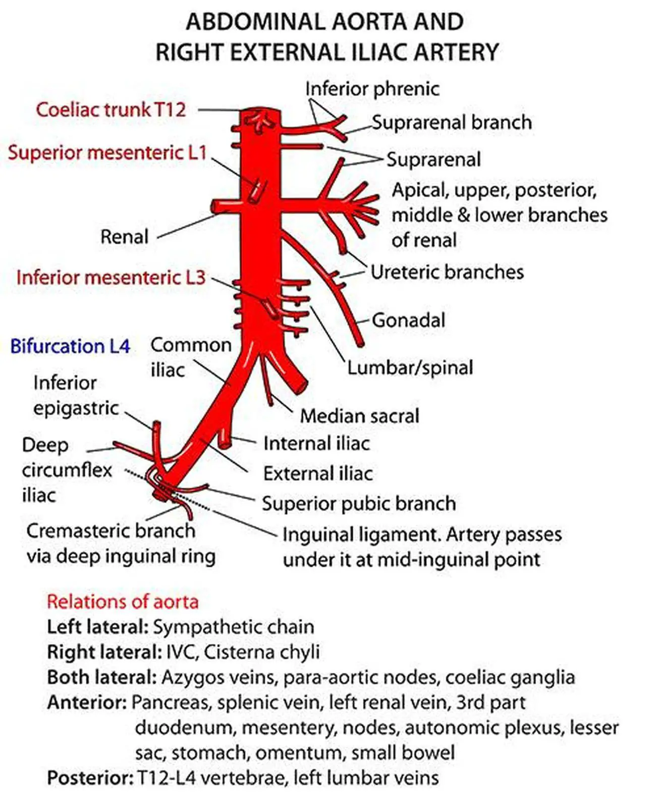
The abdominal wall surrounds the abdominal cavity, providing it with flexible coverage and protecting the internal organs from damage. The abdominal portion of the aorta supplies most of the abdomen, and begins at the level of the twelfth thoracic vertebra (t12), and then terminates at l4 by bifurcating into the left and right common iliac arteries. The diaphragm is its upper boundary. The abdominal wall is defined cranially by the xiphoid process of the sternum and the costal margins, and caudally by the iliac and pubic bones of the pelvis. The region occupied by the abdomen is called the abdominal cavity, and is enclosed by the abdominal muscles at front and to the sides, and by part of the vertebral column at the back. The regions occupied by stomach are epigastric, umbilical and hypochondriac regions. The abdominal cavity is the part of the body that houses the stomach, liver, pancreas, kidneys, gallbladder, spleen, and the large and small intestines. The normal anatomy or organs imaged in a standard abdominal examination is explained below. Abdomen, in human anatomy, the body cavity lying between the chest or thorax above and the pelvis below and from the spine in the back to the wall of abdominal muscles in the front. By convention, the abdominal exam is performed with the provider standing on the patient's right side. These two apertures, together with abdominal walls, bound the abdominal cavity. Together, these three turn nutrients into usable energy, as well as help dispose of solid waste. Together, these three turn nutrients into usable energy, as well as help dispose of solid waste.
The region occupied by the abdomen is called the abdominal cavity, and is enclosed by the abdominal muscles at front and to the sides, and by part of the vertebral column at the back. By convention, the abdominal exam is performed with the provider standing on the patient's right side. If you plan to enter a healthcare profession such as nursing, this is something you'll use on the job when performing abdominal assessments (and while documenting). Abdominal anatomy includes a major element of the gastrointestinal, system, the caudal end of the oesophagus, stomach, large and small intestine, liver, pancreas and the gallbladder. The abdominal wall is defined cranially by the xiphoid process of the sternum and the costal margins, and caudally by the iliac and pubic bones of the pelvis.
Pictures Of Abdominal Aorta from healthiack.com The diaphragm is its upper boundary. The abdomen is the part of the body that contains all of the structures between the thorax (chest) and the pelvis, and is separated from the thorax via the diaphragm. Inferiorly the abdomen is open to the pelvis, communicating through the superior pelvic aperture (pelvic inlet). External oblique, internal oblique, and transverse abdominis, supplemented in front on each side of the midline by rectus abdominis. Abdominal muscle, any of the muscles of the anterolateral walls of the abdominal cavity, composed of three flat muscular sheets, from without inward: We'll identify as many organs as we can, see how they fit into the. Boundaries of the abdomen (4) anterior abdominal wall (anterolateral) diaphragm (superior) The rectus abdominis connects to the xiphoid process, a bony landmark at the bottom of the sternum.
Part of the trunk between thorax and pelvis.
The majority of these organs are encased in a protective membrane termed the peritoneum. The abdomen contains all the digestive organs, including the stomach, small and large intestines, pancreas, liver, and gallbladder. The major organs of the abdomen include the small intestine, large intestine, and stomach. Much information can be gathered from simply watching the patient and looking at the abdomen. Inferiorly the abdomen is open to the pelvis, communicating through the superior pelvic aperture (pelvic inlet). The abdominal wall is defined cranially by the xiphoid process of the sternum and the costal margins, and caudally by the iliac and pubic bones of the pelvis. The abdomen is the body region found between the thorax and the pelvis. Together, these three turn nutrients into usable energy, as well as help dispose of solid waste. Topical anatomy of the abdomen. It follows the thorax or cephalothorax. By convention, the abdominal exam is performed with the provider standing on the patient's right side. The region occupied by the abdomen is called the abdominal cavity, and is enclosed by the abdominal muscles at front and to the sides, and by part of the vertebral column at the back. The abdominal wall surrounds the abdominal cavity, providing it with flexible coverage and protecting the internal organs from damage.
0 Response to "Abdominal Anatomy - Abdominal muscles | Golf Loopy - Play Your Golf Like a ... / This requires complete exposure of the region in question, which is accomplished as follows:"


0 Response to "Abdominal Anatomy - Abdominal muscles | Golf Loopy - Play Your Golf Like a ... / This requires complete exposure of the region in question, which is accomplished as follows:"
Post a Comment